The Pathophysiology
How High Blood Sugar Destroys Vision
The retina—the light-sensitive wallpaper at the back of your eye—is one of the most metabolically active tissues in the human body. To function, it demands a massive, constant supply of oxygen delivered through a vast network of microscopic blood vessels (capillaries).
If you have Type 1, Type 2, or Gestational Diabetes, chronically elevated blood glucose levels act like toxic shards of glass inside these capillaries. Over time, high blood sugar destroys the endothelial cells lining these vessels. The walls become weak, balloon outward, and eventually burst, leaking blood and inflammatory fluid directly into the retinal tissue.
The Illusion of Perfect Vision
Diabetic eye disease is silent. You can still have 20/20 vision while your retina is actively bleeding. Symptoms often appear only after severe damage has already occurred.
Early Warning Sign
Fluctuating Vision
Do your glasses work perfectly one day, but leave everything blurry the next?
When your blood sugar spikes, the natural lens inside your eye acts like a sponge, absorbing fluid and swelling. This rapid swelling changes the refractive index of the eye, making you temporarily very nearsighted. When your blood sugar drops back down, the lens deflates and your vision changes again.
If your vision fluctuates from day to day, you need your A1C and blood glucose checked immediately.
Disease Progression
The Stages of Diabetic Retinopathy
Diabetic Retinopathy (DR) progresses through distinct clinical stages. Early detection allows us to intervene before the disease escalates.
This is the early stage of the disease. The blood vessels in the retina weaken, but the eye has not yet started growing new, abnormal vessels. NPDR is graded from Mild to Severe based on clinical findings:
- Microaneurysms: Tiny, balloon-like swellings in the capillary walls. These are usually the very first visible sign of diabetic eye disease.
- Dot & Blot Hemorrhages: Deep retinal bleeding where the weakened capillaries have ruptured.
- Hard Exudates: Yellow deposits of lipids and proteins left behind when fluid leaks from the vessels and subsequently dries up in the retinal tissue.
- Cotton Wool Spots (Ischemia): Fluffy white patches on the retina indicating areas where nerve fibers have died because the blood supply was completely choked off.
The advanced, sight-threatening stage. Because so many blood vessels have closed off during NPDR, the retina is starving for oxygen (severe ischemia). In a desperate attempt to survive, the retina secretes a growth factor (VEGF) that forces the eye to grow brand new blood vessels.
- Neovascularization: The growth of these new vessels. However, they are highly defective, fragile, and lack structural integrity.
- Vitreous Hemorrhage: The fragile new vessels easily rupture, pouring massive amounts of blood into the clear gel cavity of the eye, causing sudden, profound blindness or severe floaters.
- Tractional Retinal Detachment: As the bleeding heals, it forms scar tissue. This scar tissue shrinks and violently pulls the retina away from the back of the eye, requiring emergency surgery.
DME is the most common cause of actual vision loss in diabetic patients, and it can occur at ANY stage of the disease (even mild NPDR).
The macula is the tiny, highly specialized center of the retina responsible for your sharp, central, reading vision and facial recognition. If the damaged capillaries leak fluid directly into the macula, the tissue swells like a blister. This swelling (edema) physically warps the photoreceptors, severely distorting and blurring your central vision.
Modern Medical Management
New Medications: The GLP-1 Effect
With the massive rise in popularity of GLP-1 receptor agonists (such as Ozempic®, Wegovy®, and Mounjaro®) for diabetes management and weight loss, understanding your ocular risk is more critical than ever.
While these medications are incredible for systemic health, they can cause a rapid, steep drop in blood sugar. Paradoxically, rapidly lowering your A1C can cause a temporary but severe worsening of existing diabetic retinopathy. The delicate blood vessels in the eye need time to adapt to new, lower glucose levels.
The NAION Risk
Recent clinical studies have also shown a potential link between semaglutide (the active ingredient in Ozempic/Wegovy) and a rare but serious condition called NAION (an "eye stroke" affecting the optic nerve). If you are starting a GLP-1 medication, a baseline comprehensive eye exam is absolutely essential.
Understanding Your Overall Risk
Diabetic retinopathy is the leading cause of blindness in working-age adults. Your risk of developing blinding complications increases based on three main factors:
Disease Duration
The longer you have had diabetes, the higher your risk of retinal damage. After 15 years, the majority of diabetic patients will show some signs of retinopathy.
A1C Levels
Chronically high or wildly fluctuating blood sugar levels act as a direct toxin, systematically destroying your ocular blood vessels.
Co-Existing Conditions
High blood pressure and high cholesterol dramatically multiply the damage diabetes does to the eye, accelerating vision loss.
State-of-the-Art Diagnostics
Inside Your Diabetic Evaluation
We do not guess. Dr. Fouladian utilizes elite imaging technology to examine the microscopic layers of your retina safely and comfortably.
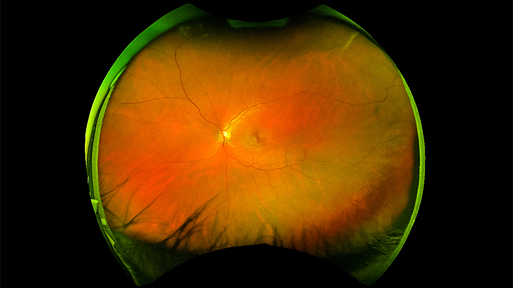
Actual Optomap® Scan
A 200° high-resolution retinal scan revealing peripheral diabetic hemorrhages often missed in standard exams.
Optomap? Ultra-Wide Imaging
A traditional exam only sees 15% of your retina at one time. Diabetic bleeding often starts in the far periphery. The Optomap captures a high-resolution, 200? digital image in a quarter of a second, allowing us to instantly spot microscopic hemorrhages-often eliminating the need for dilating drops.
Non-Invasive Vascular Imaging
Traditionally, visualizing leaking blood vessels required injecting a chemical dye (fluorescein) into your arm. Using advanced separation filters on our Optomap system, we can generate a fluorescein look-a-like image completely non-invasively to check for oxygen starvation and vessel leakage.
Stereoscopic Pupil Dilation
While Optomap is incredible, a dilated fundus exam remains the gold standard for comprehensive diabetic eye care. We instill drops to widen your pupil, allowing a stereoscopic, 3D view of the optic nerve and retinal vasculature to check for fragile neovascularization.
Your Medical Team
Collaborative Management & Treatment
Managing diabetes requires a multidisciplinary team. Dr. Fouladian acts as your ocular diagnostic hub, bridging the gap between primary care and surgical specialists.
Primary Care Coordination
The absolute best treatment for diabetic retinopathy is prevention through strict systemic control. The "ABCs" of diabetes-A1C, Blood pressure, and Cholesterol-must be managed meticulously.
After every diabetic exam, Dr. Fouladian generates a detailed medical report summarizing the health of your retinas, grading any disease severity, and sends it directly to your Primary Care Physician (PCP) or Endocrinologist. This crucial, closed-loop communication helps them accurately adjust your systemic medications.
Urgent Care
Retina Specialist Intervention
If Dr. Fouladian detects Proliferative Retinopathy (neovascularization) or Macular Edema (DME), time is of the essence to save your central vision. We will fast-track an urgent referral to an elite Retina Specialist in Los Angeles for sight-saving treatments.
Modern Treatments Include:
- Anti-VEGF Injections: Medicine injected directly into the eye to stop the growth of abnormal blood vessels and rapidly dry up macular swelling.
- Laser Photocoagulation: High blood pressure and high cholesterol dramatically multiply the damage diabetes does to the eye, accelerating vision loss.
Medical Insurance vs. Vision Plans
Because diabetes is a systemic medical disease that can cause pathological damage to the eye, a diabetic retinal evaluation is billed to your Medical Insurance (e.g., Medicare, Blue Cross, Aetna, UnitedHealthcare), NOT your routine Vision Plan (like VSP or EyeMed). Standard specialist copays or deductibles determined by your medical carrier will apply for this specialized evaluation.
Stay Ahead of the Disease
Do not wait for your vision to blur. If you are diabetic, a comprehensive annual medical eye exam is essential to preserving your sight.
TEXT FOR APPT