We organize our disease management by anatomy: from the front surface of the eye (Anterior Segment) to the retina and optic nerve at the back (Posterior Segment). Early detection in both areas is vital for preserving your vision.
Anterior Segment (Front of Eye)
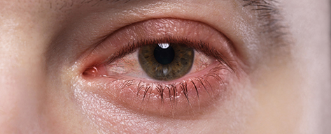
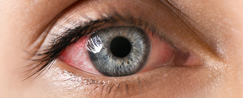
Infections (Conjunctivitis)
Viral or bacterial infections causing redness, itching, and discharge. We prescribe appropriate antibiotics or antivirals.
Diagnosis: Surface Evaluation
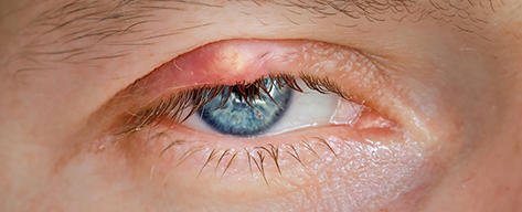
Chalazion & Stye
Lumps on the eyelid caused by blocked oil glands (Chalazion) or bacterial infection (Stye). Treatments include warm compresses or medication.
Diagnosis: Lid Examination
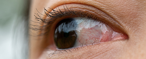
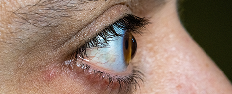
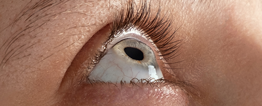
Pinguecula & Pterygium
Growths on the white of the eye often caused by UV exposure ("Surfer's Eye"). They can cause irritation and affect vision if they grow over the cornea.
Diagnosis: Slit Lamp Exam

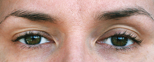
Strabismus
Misalignment of the eyes where they do not look at the same place at the same time, often affecting depth perception.
Diagnosis: Cover Test
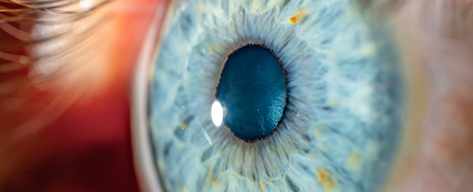
Corneal Ectasia
Progressive thinning and bulging of the cornea into a cone shape, causing irregular astigmatism and blurred vision.
Diagnosis: Corneal Topography
Narrow Angles
Anatomical crowding of the eye's drainage angle. If left untreated, it can lead to acute angle-closure glaucoma.
Diagnosis: Gonioscopy
Pseudoexfoliation
Flake-like material accumulates on the lens and drainage system, significantly increasing the risk of high eye pressure.
Diagnosis: Dilated Slit Lamp Exam
Pigmentary Dispersion
Pigment rubs off the back of the iris and clogs the eye's drainage meshwork, potentially causing Pigmentary Glaucoma.
Diagnosis: Transillumination
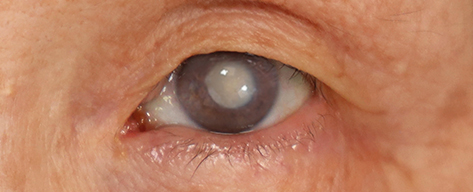
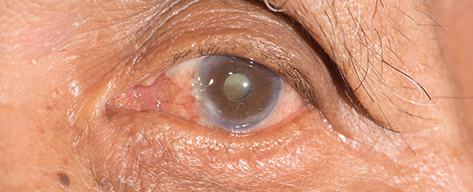
Cataract
A clouding of the eye's natural lens that leads to blurred or yellowed vision. It is a natural part of aging but can be treated surgically.
Diagnosis: Slit Lamp Exam
Uveitis & Iritis
Inflammation of the middle layer of the eye (uvea). It causes redness, pain, and light sensitivity.
Diagnosis: Slit Lamp Exam
Eyelid Ptosis
Comprehensive evaluation of drooping eyelids to protect your vision and underlying health.

Double Vision & Pupil Disorder
When visual symptoms are a warning sign of systemic or neurological disease.
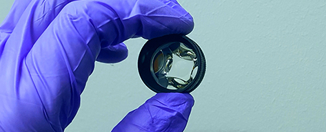
Degenerative Corneal Diseases
Advanced diagnostics, medical therapy, and specialty lens rehabilitation for complex corneal conditions.
Posterior Segment (Back of Eye)
Glaucoma
Damage to the optic nerve, often due to high pressure. Leading cause of irreversible blindness.
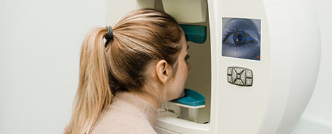
Diagnosis: OCT & Visual Field
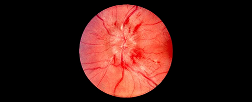
Papilledema
Swelling of the optic nerve caused by increased pressure in the brain. Requires immediate medical attention.
Diagnosis: Emergency Fundus Exam
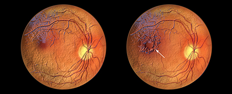
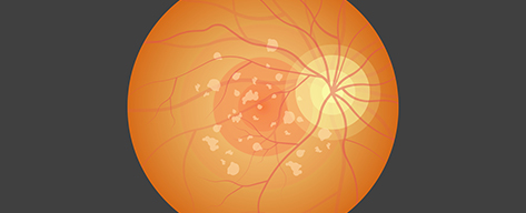
Diabetic Retinopathy
Damage to retinal blood vessels caused by high blood sugar. Can lead to leakage, swelling, and vision loss.
Diagnosis: Optomap Imaging
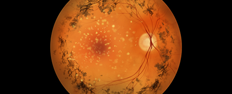
Hypertensive Retinopathy
Retinal damage caused by high blood pressure. Arteries in the eye can narrow or burst.
Diagnosis: Fundus Exam

Macular Degeneration
Deterioration of the central retina (macula), affecting your ability to see fine details and read.
Diagnosis: Dilated Exam & OCT
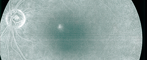
Serous Maculopathy
Fluid accumulation under the retina causing a central blur. Often triggered by stress or steroid use.
Diagnosis: OCT Imaging
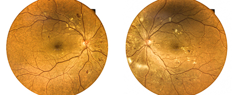
Fundus Flavimaculatus
A genetic condition (Stargardt variant) characterized by yellow-white flecks in the retina affecting central vision.
Diagnosis: Autofluorescence
Retinitis Pigmentosa
A genetic disorder causing loss of retinal cells, leading to tunnel vision and night blindness.
Diagnosis: Visual Field & ERG
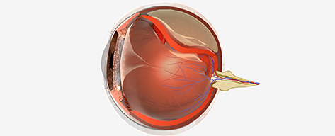
Retinal Detachment
Emergency where the retina pulls away from the eye wall. Warning signs: flashes and floaters.
Diagnosis: Emergency Dilated Exam
Early Detection Saves Sight
Do not wait for symptoms to appear. Schedule your comprehensive medical eye exam today.
TEXT FOR APPT